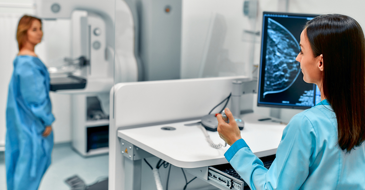
Used to perform routine breast cancer screenings, or to diagnose conditions in women who have other concerning symptoms, mammography is a type of medical imaging that captures images of breast tissue through low-dose X-rays. Since its first FDA approval in 2011, the 3D mammogram has increasingly become the preferred method in diagnostic centers across the U.S.
As it is part of our mission to provide the latest technology for comprehensive care of women’s wellness, we want to take a moment to explain some of the benefits of 3D mammography.
What Is a 3D Mammogram?
Traditional mammography uses two-dimensional imaging to look at breast tissue. 3D mammography, also known as breast tomosynthesis, is a more advanced form of this digital imaging. The series of images in 3D mammography are created by running low-dose X-rays over compressed tissue. The images are captured by a computer, which assembles them together for a comprehensive examination of glandular breast tissue.
3D Mammograms Vs. Traditional Mammography
Like a traditional mammogram, 3D breast imaging still requires the compression of breast tissue, so your experience of 3D mammography might not differ much from what you’ve felt with a traditional mammogram. Yet, from the perspective of experts like Cynthia A. Litwer, MD, chief of breast imaging at Cedars Sinai, and those at Yale Medicine, 3D images seem to provide improved diagnostic results.
The reason? Two-dimensional imaging captures pictures of breast tissue from four limited angles — such as the top or side. Tumors and other abnormalities can therefore get hidden in these flat, overlapping images. In 3D mammography, however, the imaging machine moves along an arc trajectory, capturing 200 to 300 images in total.
If a 2D mammogram fails to provide adequate answers, patients must go for subsequent imaging (including, possibly, a breast ultrasound) for an accurate diagnosis. Because the 3D imaging process provides more images in greater detail, it can reduce the necessity of potentially stressful call-backs for follow-up images.
Using 3D mammography could also be more effective for women with dense breast tissue. In addition to milk ducts, glands, and fatty tissue, breasts are also made up of supportive, connective tissues. In areas of this tissue density, pinpointing troubling anomalies can be challenging. But the improved imaging of 3D mammography provides a much clearer, comprehensive picture of what’s going on — all in a single screening.
Increasing The Chances of Treatment and Remission
While there is ongoing research comparing clinical outcomes between the two approaches, 3D mammography’s more extensive imaging could detect more cases of breast cancer in the earliest stages. As the exact causes of breast cancer still remain unknown, experts at the World Health Organization assert, “early detection of the disease remains the cornerstone of breast cancer control.”
Mammograms can help doctors diagnose cancer before patients feel any lumps or abnormalities. And lumps detected with 3D mammography, according to a 2019 study in JAMA Oncology, “were more likely to be smaller and node-negative (meaning they had not spread to the lymph nodes) compared to breast cancers found with standard digital mammography.” Meaning: the cancers were detected before they were able to worsen and spread.
As a result, 3D mammography may be saving even more lives than its 2D sister technology.
Whether you’re due for a breast cancer screening, or your doctor has prescribed a diagnostic mammogram, let the caring team at Cardinal Points Imaging provide first-rate care. To schedule your 3D mammogram — or another imaging test — call (919) 877-5400 or contact us online.

